— Life on Earth —
Our mission is to create a shared plan
for saving the planet’s most exceptional wild places.
ABOUT US
Shaping our future
With all the global problems our planet faces today,
communities of people concerned with them are growing
to prevent the negative impact.

Changing the world is possible.
We’ve done it before.
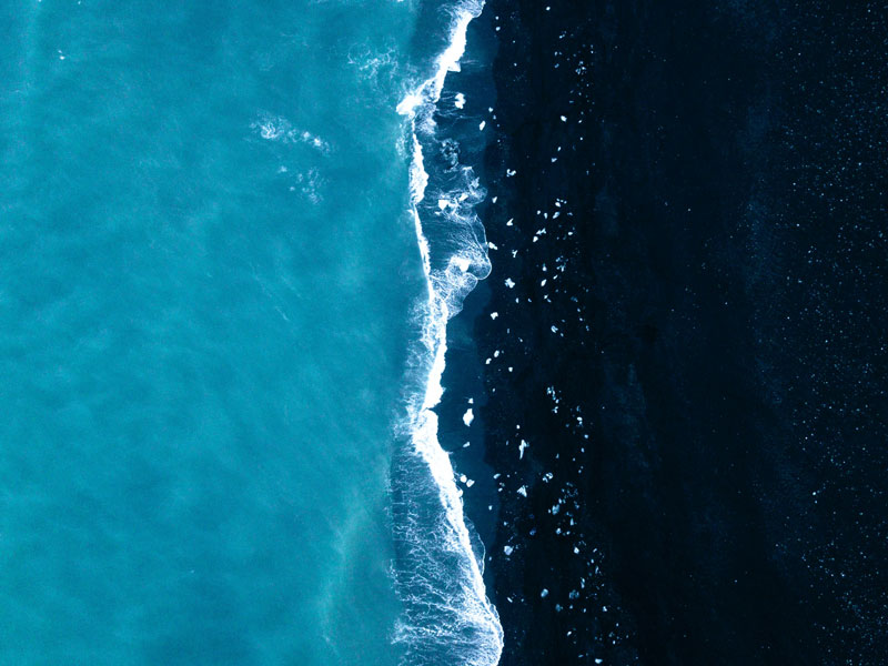
Recycling water
Recycling water for reuse applications instead of using freshwater supplies can be a water-saving measure.
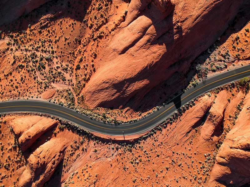
Habitat Model
Habitats with a minimum footprint on the planet and a maximum positive impact on the local community.
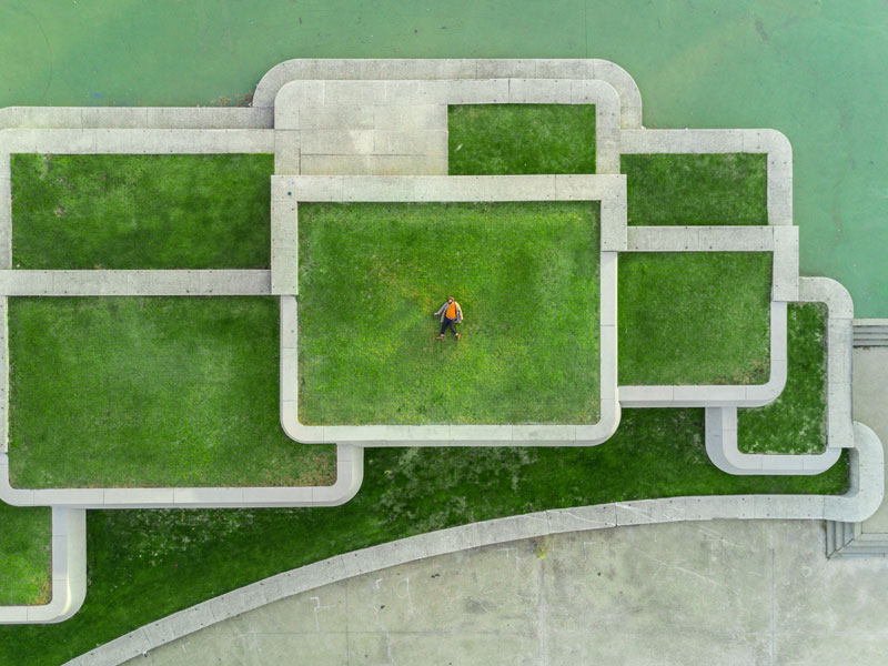
Organic Garden
Learn how to use organic gardening methods to grow the freshest food in your fruit and vegetable garden.
Help us protect and preserve for future generations
Join us and make the planet a better place.